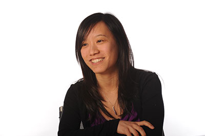
Hometown, Country:
Walnut, California, USA
Academic history prior to coming to MIT:
B.Sc. in Electrical Engineering and B.Sc. in Biological Sciences, Stanford University
What brought you to MIT?
Boston is a premier site for biomedical research in the US. There are strong collaborations between academic researchers, investigators at research centers such as the Athinoula A. Martinos Center for Biomedical Imaging, and clinicians at world-renowned hospitals in the area. The EECS department at MIT encourages interdisciplinary work and provided me the ideal environment to develop imaging technology and translate our new methods to the clinic. Also, this California girl was curious to experience “seasons” on the East Coast (she had heard so much about them!).
What problem are you trying to solve with your current research and what are some possible applications?
Our work aims to create new MRI technology to image oxygen consumption in the human brain. The healthy brain consumes 20% of total body energy through aerobic metabolism, and continuous oxygen delivery is necessary to maintain the health of neural tissues. Unfortunately, this oxygen supply is disturbed in many neurological disorders, including stroke, tumor, and multiple sclerosis. The ability to image oxygenation would help clinicians diagnose early metabolic changes in these diseases and select the appropriate therapy for patients. However, there is currently no clinically feasible way to measure oxygen consumption the brain. We aim to provide a new imaging tool for radiologists to directly read oxygenation levels across different regions of the brain.
What interests you most about your research?
I love being able to interact with scientists and clinicians of very diverse backgrounds. My research group, for instance, has strong expertise in signal processing and optimization, as well as in design and implementation of new MRI pulse sequences. Their guidance has been instrumental in my method development work. In addition, I’ve had the opportunity to collaborate with clinicians such as Dr. Bruce Rosen and Dr. Caterina Mainero at the Martinos Imaging Center to study patients with multiple sclerosis. Their clinical needs have provided valuable directions to refine our methods for eventual use in hospitals.
What are your future plans?
In the future, I see myself as a faculty member or investigator who can work closely with engineers and doctors to develop new radiological tools for hospitals. I also really enjoyed my experiences as a TA at MIT and hope to teach in some capacity for my future career.
Learn more about the Magnetic Resonance Imaging Group
